|
PinkMonkey Online Study Guide-Biology
(B) Female reproductive system (Fig. 24.4)
The female reproductive system consists of following parts:
(1) Ovaries.
The ovaries are two oval bodies (about 30-40 mm long) located below and
behind the uterine tubes. Each ovary is supported by a series of ligaments.
The main function of the ovary is to produce eggs (ova) and the female
sex hormones, estrogen and progesterone. They are attached
by a membrane to the uterus and supplied with blood vessels.
(2) The Uterine Tubes.
The uterine tubes consist of the proximal
part called oviducts and the distal part called Fallopian tubes.
The uterine tubes are attached to the uterus at its upper outer angles.
The tubes are lined with cilia and extend to the ovaries. At the distal
end each fallopian tube expands into funnel like infundibulum,
having the fringe like projections called fimbriae. When ova are
released from the ovary they pass down the fallopian tube. Fertilization
normally occurs in the Fallopian tube.

Figure 24.4 Female reproductive system
(3) The Uterus.
Both oviducts open into a wider tube, the uterus, or womb, which measures
about 3 inches in length, 2 inches in width and 1 inch in depth. The uterus
consists of a dome-shaped portion, called the fundus, a central tapering
portion- the body and a narrow portion opening into the vagina, called
the cervix.
(4) The vagina is a collapsible muscular tube, about 3 inches long, and capable of great distention. At the lower end of the vagina normally there is a fold of mucous membrane in the virginal state, the hymen, which partially closes the orifice. The vagina receives the seminal fluid, serves as the lower part of the birth canal and acts as an excretory duct for uterine secretions and menstrual flow.
(5) The vulva
is a collective name for female external genitalia. Two folds of skin,
the labia majora and the labia minor, enclose the vulva
at the entrance to the vagina.
(6) The Bartholinís or vestibular
glands are present on either side of
the vaginal orifice, and secrete a lubricating fluid.
(7) The mammary glands,
or breasts, are functionally related to the female reproductive system,
since they secrete milk for the nourishment of the young.
A transverse section of an ovary (Fig-24.5A) of ovary
shows that it consists of an outer cortex and inner medulla.
The medulla contains the connective tissue called stroma, blood
vessels and nerve fibers. The cortex is lined by germinal epithelium,
below which there are groups of follicles, each enclosing an egg.
The cortex is lined externally by a dense fibrous tissue,
the tunica albuginea. Out of many follicles present at birth, only 300
to 400 follicles reach maturity and liberate their ova (ovulation)
during the reproductive years of a human female (puberty to menopause).
One of the developing follicles (primary follicle, secondary follicle,
maturing follicle) form a mature follicle called the Graafian follicle.
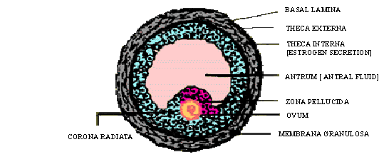
Each Graafian follicle contains the ovum. Immediately
surrounding the ovum is a layer of follicle cells, the inner zona pellucida
and outer layer of columnar cells, the corona radiata. The follicle
contains a fluid filled space called the antrum. The antrum is
lined by follicle cells which from two zones, the membrana granulosa
and the cumulus oophorus. The Graafian follicle is surrounded by
two connective tissue cell layers, the outer theca externa and
the inner theca interna. The follicular cells secrete the hormone
estrogen which controls the menstrual cycle.

click here for enlargement
Figure 24.5 (A) Transverse section through an Ovary

click here for enlargement
Figure 24.5 (B) Graafian follicle
[next page]
|