Figure 22.5 Thyroid gland
(A) Position and Structure: All vertebrates
possess a pair of thyroid glands, located in the neck just
below the larynx or Adams apple. In humans, the thyroid consists
of two lobes (H-shaped) that lie on either side of the trachea.
The two lobes are connected by a narrow isthmus that passes in front
of the trachea (Figure 22.5). The gland varies in size with the
difference in sexual development, diet and age.
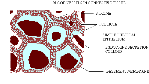
(B) Histology. The thyroid gland consists
of a large number of round or oval follicles surrounded by
connective tissue with a large number of blood vessels. Each follicle
is lined by one-cell thick cuboidal epithelial cells. The
cavities of the follicles are filled with viscous protein material
called colloid (Figure 22.6). The thyroid hormone, thyroxine
contains iodine atoms. It is not stored in the cells of the thyroid,
but in the colloid filling the follicles. The role of thyroid cells
is to strain iodine out of the blood. This is then incorporated
into the protein thyroglobulin, which is then hydrolyzed
into the active hormone thyroxine.

Click here for enlargement
Figure 22.6 Transverse section through the thyroid
(C) Thyroid Hormones
As mentioned earlier, thyroid glands produce a
hormone called thyroxine. The secretion of thyroxin is controlled
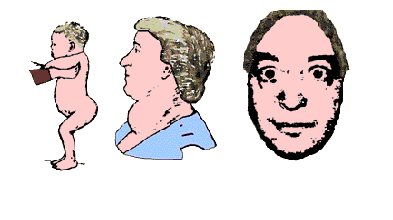
by TSH produced by the pituitary. The hyposecretion of thyroxine
in a child results in cretinism (Figure 22.7 A). In this,
retarded growth and development (dwarfism), a protruding abdomen,
mental retardation, puffy skin and low metabolic rate occurs.

(A)Cretinism (B)Goiter (C)Exopthalmos
Figure 22.7